
A new year — and decade — offers the opportunity to reflect on the advancements and challenges of years gone by and ...
Cardiac Imaging
The cardiac imaging channel includes the modalities of computed tomography (CT), cardiac ultrasound (echocardiography), magnetic resonance imaging (MRI), nuclear imaging (PET and SPECT), and angiography.

March 28, 2020 — Women with coronary artery disease that reduces blood flow and oxygen to the heart muscle (ischemia) ...
 March 28, 2020
March 27, 2020 — In Italy today, novel coronavirus (COVID-19) claimed the life of well known cardiologist Maurizio ...
March 27, 2020Sponsored Content

Feature
SPONSORED CONTENT — Studycast is a comprehensive imaging workflow system that allows healthcare professionals to work ...
June 17, 2025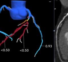
James Udelson, M.D., chief of the division of cardiology, Tufts Medical Center, explains how cardiac computed tomography ...
March 26, 2020
Interview with Mike Stone, M.D., an emergency physician at Northwest Acute Care Specialists in Portland, Ore., director ...
March 17, 2020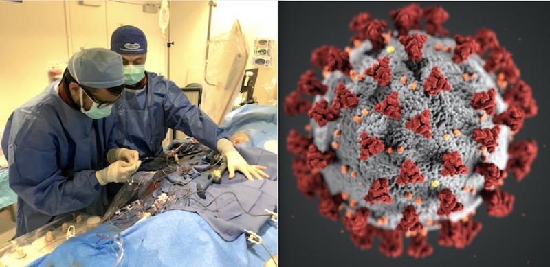
Feature | Coronavirus (COVID-19) | Dave Fornell, Editor
March 20, 2020 — The Centers for Medicare and Medicaid Services (CMS) announced March 18, 2020, that all elective ...
March 20, 2020Sponsored Content
Feature | Cardiac Imaging
Cardiac positron emission tomography (PET) is growing in popularity among cardiologists because it provides the ability ...
March 05, 2024
An interview with Ehtisham Mahmud, M.D., FSCAI, chief, Division of Cardiovascular Medicine, executive director of ...
March 20, 2020
March 16, 2020 — The Society of Cardiovascular Computed Tomography (SCCT) released a new expert consensus document on ...
March 16, 2020
Feature | Angiography | Dave Fornell, Editor
Philips is working on a prototype cath lab angiographic imaging system that might be able to replace the current X-ray ...
March 12, 2020Sponsored Content

Blog | Enterprise Imaging
As medical advancements continue to push the boundaries of what is possible in the field of structural heart ...
August 10, 2023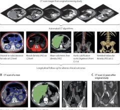
March 6, 2020 — Researchers at the National Institutes of Health and the University of Wisconsin have demonstrated that ...
March 06, 2020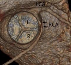
March 5, 2020 — Experts in the medical imaging community have developed a new, landmark consensus document to optimize ...
March 05, 2020
March 5, 2020 — More than 300 patients have joined the Amyloidosis Patient Registry and it is now available to the ...
March 05, 2020Sponsored Content

Feature | Cardiac Imaging
Discover the key features of cardiovascular structured reporting that drive adoption, including automated data flow, EHR ...
November 07, 2022
Feature | Dave Fornell, Editor
March 3, 2020 — Two sizable European medical conferences in cardiology and radiology were canceled today as fears ...
March 03, 2020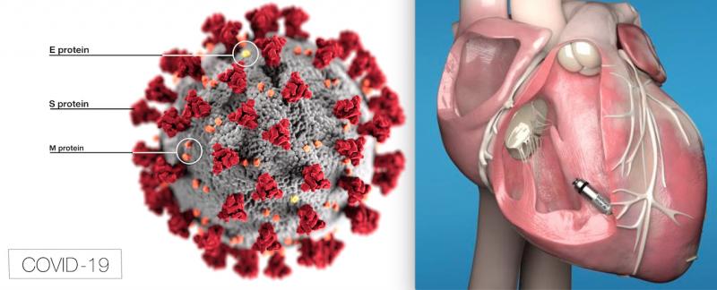
Feature | Dave Fornell, Editor
March 2, 2020 — Here is the list of the most popular content on the Diagnostic and Interventional Cardiology (DAIC) maga ...
March 02, 2020
February 28, 2020 — New healthcare technologies are being implemented in the fight against the novel coronavirus (COVID ...
February 28, 2020