October 1, 2020 — The U.S. Food and Drug Administration (FDA) granted 510(k) clearance for the SentiAR CommandEP system ...
Cardiac Imaging
The cardiac imaging channel includes the modalities of computed tomography (CT), cardiac ultrasound (echocardiography), magnetic resonance imaging (MRI), nuclear imaging (PET and SPECT), and angiography.

 October 01, 2020
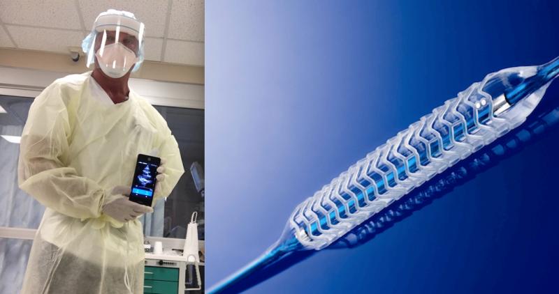
Feature | Dave Fornell, Editor
October 1, 2020 — Here is the list of the most popular content on the Diagnostic and Interventional Cardiology (DAIC) ...
October 01, 2020
Feature | Artificial Intelligence | Joe Fornadel, J.D., and Wes Moran, J.D.
The number of Federal Drug Administration (FDA)-approved AI-based algorithms is significant and has grown at a steady ...
September 29, 2020Sponsored Content

Feature
SPONSORED CONTENT — Studycast is a comprehensive imaging workflow system that allows healthcare professionals to work ...
June 17, 2025
Ernest Garcia, Ph.D., MASNC, FAHA, endowed professor in cardiac imaging, director of nuclear cardiology R&D laboratory ...
September 25, 2020
September 25, 2020 — Based on its recent analysis of the global artificial intelligence (AI)-based echocardiography mark ...
September 25, 2020
September 25, 2020 — A study out of University Hospitals (UH) found that removing the cost barrier for coronary artery ...
September 25, 2020Sponsored Content
Feature | Cardiac Imaging
Cardiac positron emission tomography (PET) is growing in popularity among cardiologists because it provides the ability ...
March 05, 2024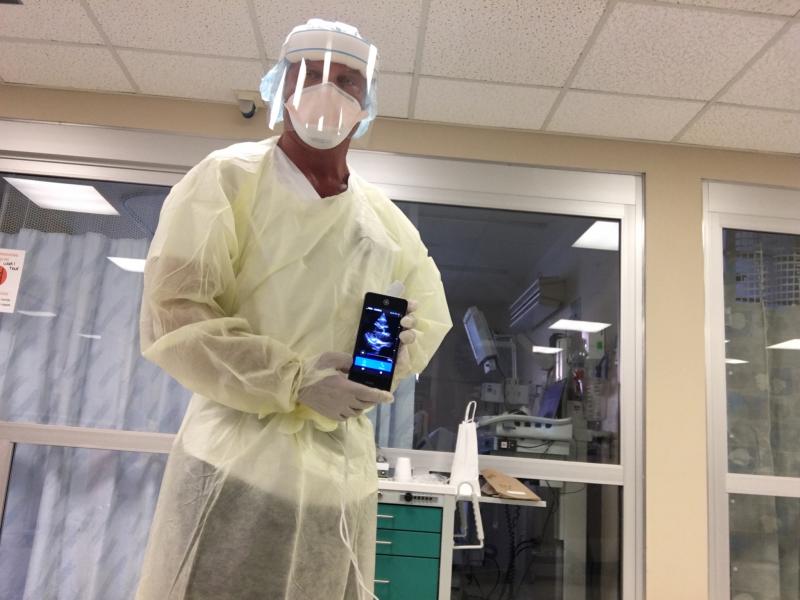
September 15, 2020 — The world is facing a global pandemic with unknown implications, but is now well known COVID-19 ...
September 15, 2020
Case is a 6-month-old infant boy admitted to hospital due to respiratory distress then worsened by a pericardial ...
September 15, 2020
September 15, 2020 — Philips Healthcare recently introduced the latest addition to its portfolio of dedicated cardiovasc ...
September 15, 2020Sponsored Content
Blog | Enterprise Imaging
As medical advancements continue to push the boundaries of what is possible in the field of structural heart ...
August 10, 2023
Matthew Budoff, M.D., director of cardiovascular CT at The Lundquist Institute, and professor of medicine at the David ...
September 10, 2020
September 10, 2020 - Icosapent ethyl (Vascepa) demonstrated significant, 17 percent regression of low attenuation plaque ...
September 10, 2020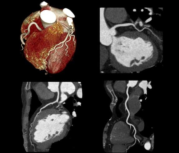
September 8, 2020 — The Journal of the American College of Cardiology (JACC) published a report, “Current Evidence and ...
September 08, 2020Sponsored Content

Feature | Cardiac Imaging
Discover the key features of cardiovascular structured reporting that drive adoption, including automated data flow, EHR ...
November 07, 2022
September 2, 2020 — Patient-specific organ models are being used by the University of Minnesota to better prepare for ...
September 02, 2020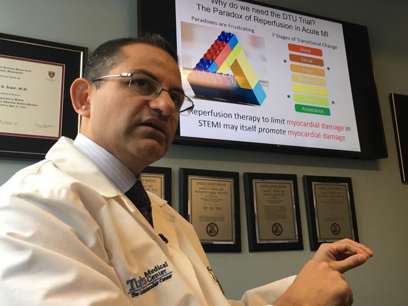
Feature | Tufts Medical Center | Dave Fornell, Editor
The cardiology program at Tufts Medical Center in Boston is internationally recognized for being on the forefront of ...
August 21, 2020
August 20, 2020 – More than 350 posters featuring cutting edge research on the advances in cardiovascular ultrasound ...
August 20, 2020