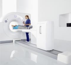
June 15, 2022 — A cardiac SPECT imaging system performs scans 10 to 100 times faster than current SPECT systems ...
Cardiac Imaging
The cardiac imaging channel includes the modalities of computed tomography (CT), cardiac ultrasound (echocardiography), magnetic resonance imaging (MRI), nuclear imaging (PET and SPECT), and angiography.
June 15, 2022 — Poor functional outcomes after a heart attack can be predicted with a new PET imaging agent, 68Ga-FAPI ...
June 15, 2022 — Six radiology and cardiology trainees have been selected as finalists for the 16th Annual Canon Young ...
SPONSORED CONTENT — Studycast is a comprehensive imaging workflow system that allows healthcare professionals to work ...
The X5-1c transducer from Philips provides enhanced clinical information in transthoracic imaging over a standard phased ...
June 14, 2022 — A new American Heart Association scientific statement highlights the need for more data and research ...
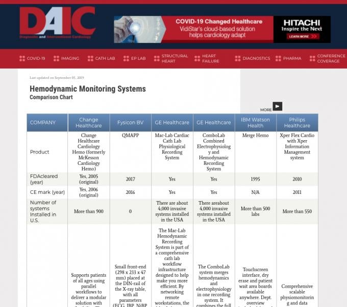
Diagnostic and Interventional Cardiology (DAIC) maintains more than 50 comparison charts of product specifications from ...
Cardiac positron emission tomography (PET) is growing in popularity among cardiologists because it provides the ability ...
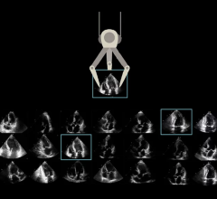
June 8, 2022 — Us2.ai, a Singapore-based medtech firm backed by, IHH Healthcare, Heal Partners, Sequoia India, EDBI ...
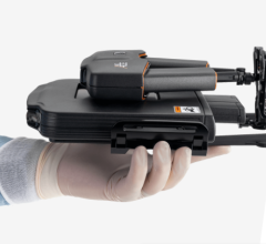
June 7, 2022 — Visura Technologies, Inc., a privately-held medical device company dedicated to delivering state-of-the ...
June 7, 2022 — ScImage Inc., a leading provider of enterprise imaging solutions and DiA Imaging Analysis, a global ...
As medical advancements continue to push the boundaries of what is possible in the field of structural heart ...
May 31, 2022 — Innovative Health, LLC, a specialty cardiology reprocessor, announced that the company has received ...
May 27, 2022 — Medtronic today announced approval from the U.S. Food and Drug Administration (FDA) for the IN.PACT 018 ...
Discover the key features of cardiovascular structured reporting that drive adoption, including automated data flow, EHR ...
May 26, 2022 — At the 69th Annual Conference of the Israel Heart Society, UltraSight, an Israeli-based digital health ...
May 19, 2022 — One year outcomes from the Disrupt PAD III Trial comparing intravascular lithotripsy (IVL) with a drug ...
May 18, 2022 — XACT Robotics, developer of the world’s first and only comprehensive robotic system for interventional ...


 June 15, 2022


