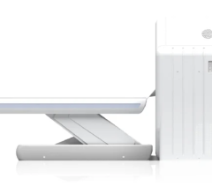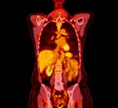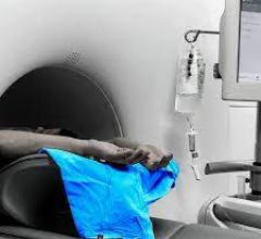
The Biograph mCT PET/CT scanner enables facilities to serve both the nuclear medicine and the radiology departments with one system. It offers CT configurations up to 128 slices. Siemens also offers the Symbia family of SPECT and SPECT/CT scanners.
Technology in the field of nuclear cardiac imaging — single photon emission computed tomography (SPECT) and positron emission tomography (PET) — has been somewhat stagnant over the past decade. However, there are some recent advancements in both radiotracers and hardware that could change this field of imaging.
“This is a very exciting time in nuclear cardiology,” said Diwakar Jain, M.D., FACC, FRCP, FASNC, professor of medicine, director of nuclear cardiology, Drexel University College of Medicine. He spoke to Diagnostic & Interventional Cardiology magazine to highlight several advancements in the field of cardiac nuclear imaging.
Improved SPECT Cameras
New gamma cameras entering the market are much faster and offer better quality images than previous versions. Three are currently on the market that use solid-state, cesium iodide detectors that directly convert gamma rays into electrical impulses, instead of traditional sodium iodide detectors that first convert the rays into photons that are then converted into electrical signals. This reduces a step in the image generation process and increases the quality of the image.
“These cameras are much better quality and have better resolution. They are somewhat new and expensive, but with time they will become more affordable,” Jain said.
Heart Failure SPECT Imaging
A new radiotracer, iodine 123, (AdreView) is being developed by GE Healthcare to image the sympathetic nervous system to help improve heart failure (HF) screenings. HF impairs uptake of this agent.
Jain said HF is usually graded based on ejection fractions, but there is a lot of overlap between the severity of the different New York Heart Association HF classes. He said the new agent may offer a more accurate stratification of HF.
The tracer is expected to have a big impact on determining which HF patients should receive an implantable cardioverter defibrillator (ICD) or cardiac resynchronization therapy (CRT) device. Jain said only about 10 percent of these are ever activated in HF patients, but are widely implanted as a precaution. He said the iodine 123 may help identify which patients will actually benefit from these devices.
Phase III clinical data have been submitted to the FDA and it is possible the tracer could receive FDA clearance by the end of 2010. If cleared, Jain said it would be the first new SPECT radiotracer cleared by the FDA since Myoview in 1994.
Results from the ADMIRE-HF (AdreView Myocardial Imaging for Risk Evaluation in Heart Failure) trial were published in the May 2010 issue of the Journal of the American College of Cardiology. The prospective study evaluated cardiac sympathetic nerve imaging using the AdreView. According to the results, in appropriately selected patients with heart failure, the imaging procedure can alert clinicians to the potential need to consider additional treatments.
Hybrid Imaging
Combined SPECT/computed tomography (CT) imaging systems are being used for better attenuation to create better images. The CT data also offer an anatomical roadmap to pinpoint specific anatomy and match it to the nuclear images.
Theses hybrid systems remain pricey, but have the advantage of being used as standard CT scanners when SPECT imaging is not being performed to increase room and device utilization.
Isotope Shortage Aids PET
PET Imaging offers better image quality and faster scanning times than SPECT, but Jain said it is not widely used in cardiac imaging. The main reason is PET is more expensive and the isotopes have such a fast decay rate they must be created onsite using a cyclotron, which greatly limits their availability.
Jain said one boost to PET in the past year is due to the SPECT isotope shortage caused by the shutdown of the Chalk River Reactor in Canada. He said the reactor is back online, but as with all the medical isotope reactors in operation around the world, it is old. All of these reactors will have periodic shutdowns for an increasing amount of maintenance.
PET can use four or five different radiotracers. The standard one used in cardiac imaging is rubidium 82 (CardioGen 82).
“The biggest issue with rubidium is the cost – it is very, very expensive, $30,000 to $40,000 per month,” Jain said. “Rubidium 82 only has one use — for cardiology, but it does not make sense with lower volume labs.”
The short 75-second half-life of the radiotracer is also a drawback. Jain said it forces use of pharmacologic stress rather than the preferred method of exercise stress.
Other isotopes can be used for cardiac imaging, including oxygen 15 and nitrogen 13, but they have not been commercialized and also require production in an on-site cyclotron because of a short 10-minute half-life, Jain said. Cyclotrons are usually only available at research facilities or at extremely high-volume labs that image 30-40 patients a day to make the massive hardware investment feasible.
“Radiotracers are the limiting factor with PET,” Jain explained.
Deficit Reduction Act Reverses Trend
A few years ago the trend in cardiac nuclear imaging was toward smaller, less-expensive cameras aimed at expanding office-based imaging. However, with the Centers for Medicare and Medicaid (CMS) instituting the Deficit Reduction Act (DRA) a couple of years ago, the brakes were put on office-based imaging. The act instituted a 30 percent cut in office-based imaging, because of the CMS perception that many of these self-referrals are motivated by revenue rather than patient diagnostics.
New PET Agents
Currently in trial is a new fluorine 18 (F18) PET tracer developed by Lantheus that has a two-hour half-life, allowing it to be produced at a commercial cyclotron and transported to numerous hospitals in the area. Lantheus presented phase II data at the 2010 Society of Nuclear Medicine (SNM) annual meeting for its flurpiridaz F18 injection. The data showed better image quality and an increase in defect size compared to SPECT.
“The results have been very promising,” Jain said. “It has all the benefits of PET imaging, which is inherently better than SPECT imaging.”
He said there is a reasonable chance the FDA may consider approval for the agent in 2011.
Another tracer that could potentially make cardiac PET imaging more accessible and widespread is gallium 68. Jain said it has the advantage of the parent isotope, germanium 68, having a 271-day half-life. This allows it to be shipped to imaging facilities in a generator and allows long-term use. Once activated, the gallium 68 isotope has a half-life of about 68 minutes.
“This would clearly be a big development,” Jain said.
Potential generator costs of between $12,000 to $20,000 a year would definitely be a price break advantage over a rubidium 82 generator costing between $30,000 to $40,000 per month.
Direct Ischemic Imaging
Another innovation being researched by Jain is myocardial ischemia imaging to directly identify damaged myocardium, rather than imaging for evidence of ischemia. He said ATP, usually in the form of free fatty acid, is generally used by healthy heart muscle in the presence of oxygen. However, when oxygen is not available, as in ischemic tissue, the muscle can convert glucose into energy. Jain said deoxiglucose (FDG) can be used as a biomarker


 November 17, 2025


![Phase III clinical trial of [18F]flurpiridaz PET diagnostic radiopharmaceutical meets co-primary endpoints for detecting Coronary Artery Disease (CAD)](/sites/default/files/styles/content_feed_medium/public/Screen%20Shot%202022-09-13%20at%203.30.13%20PM.png?itok=2w6OoNd6)
