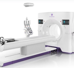
Doctors can walk up to and through an MRI or CT scan, or even
Photos of medical images produced from a new 3-D volume rendering software have the appearance of something from a “Star Trek” episode, but this virtual reality system is 100 percent reality and heading for the European healthcare market place.
Scientists at Erasmus University Medical Center, Rotterdam, The Netherlands, have developed I-space, which is capable of converting 2-D medical images acquired from conventional imaging modalities (such as MR, CT and ultrasound) into 3-D images with which doctors can truly interact. For the commercial launch of the I-space model, the medical center chose Barco projectors and SGI's Silicon Graphics Prism visualization systems as image generators to create the four-sided, 3-by-3-by-3 meter, 3-D images, according to a press release.
Crosslinks, the company created by the Erasmus University MC, is beginning to market I-space to hospitals in Europe in collaboration with SGI and Barco.
“We built I-space ourselves in collaboration with the folks from SGI and Barco,” said I-space inventor professor Dr. Peter van der Spek, a geneticist by training who is also an engineer. “Barco has the projectors, but the Silicon Graphics Prism has all the graphics pipes that superbly work together with Barco. It's cutting-edge technology and the power of Intel inside the Prism system is also very important for us, so we are very, very happy with the technology that drives I-space.”
Providing multiple clinical applications in which physicians can literally walk up to and through still and real-time images, I-space offers the important opportunity for cardiologists to examine cardiac infarction — scrutinizing live, beating hearts within I-space to see which part of the heart is paralyzed due to an infarct.
“It's easier to make a diagnosis together with other medical personnel when the surgical reality is right in front of you, rather than seeing it alone, just on a small computer screen,” said Ronald Nanninga, founder and managing director of Crosslinks.
Technically Ingenious
A Silicon Graphics Prism visualization system with eight Intel Itanium 2 processors, eight ATI FireGL graphics processors and 12GB of memory is used to drive I-space, which uses eight Barco projectors for the four walls: the floor, and the left, right and front.
A 3-D mouse uses four tracking devices — one in each corner, enabling the system to recognize the relative position of the mouse — and a virtual stick, which allows the user to touch an object, push it on one side, zoom in/zoom out and even slice the object. Stereoscopic glasses complete the immersive 3-D experience.
“We can do all kinds of very sharp visualizations, which in turn allow us to do very precise measurements on the MRI scans, on CT scans or on ultrasound images. We can zoom in really close, within the scan, to make it larger...” said Dr. van der Spek. “The very strong graphics capacity of the SGI equipment, including its software and the OpenGL graphics libraries, allows us to visualize, really, life in 3-D.”


 February 02, 2026


