Vendors showcase the latest medical imaging technological advances each year during the annual Radiological Society of North America (RSNA) meeting in Chicago, always held the week following Thanksgiving. After spending a week walking the show floor and meeting with scores of vendors at RSNA 2011, the following are my choices for the most innovative new technologies presented this year.
A New Chapter Opens for Ultrasound Visualization
Toshiba introduced its Aplio 500 ultrasound system, which takes 3-D ultrasound to a new level. It offers a fly-through feature so users can follow a centerline cine view of the interior of vascular or other hollow liquid-filled structures.
The image quality is similar to computed tomography (CT) 3-D fly-through reconstruction, as seen in virtual colonoscopy. To the untrained eye, most would think the fly-through images were from CT, so this is a big leap forward for ultrasound in terms of image quality and technical ability. When I first began covering medicine five years ago, I saw a CT coronary artery fly-through reconstructed from a CT dataset. The researcher/presenter said it took about 40 hours of computing time to create the short cine loop and said it would be years before the technology could be commercialized. Today, it can be done in minutes, it is being commercialized and it can be done by ultrasound without ionizing radiation.
The Aplio 500 also has a feature where a CT dataset can be synchronized with the ultrasound image in a side-by-side display for improved procedural navigation. As the transducer is moved, the CT dataset will react to show the exact same image on the opposite screen.
A New Breed of Angiography Systems
GE introduced a new type of angiography system that offers the image quality of a fixed ceiling or floor-mount system, but with the mobility of a mobile C-arm. The IGS 730 is basically a fixed angiography system mounted on wheels so it can move anywhere in a cath lab or hybrid OR. It uses a laser locator system to precisely align the system with the patient table. This enables high-quality images, precise gantry positioning, rotational angiography and advanced imaging features, such as image overlays, as found on ceiling or floor-mounted systems.
As it moves about the room, the system can detect obstacles, such as power cords, and has a device to gently sweep them out of the way. It also has anti-collision detectors to prevent the system from running into equipment, booms or monitors.
The IGS 730 is under regulatory review in both Europe and the United States.
MRI to Replace Angiography?
Philips Healthcare showed conceptual work-in-progress technology where magnetic resonance imaging (MRI) could be used in an interventional/cath lab suite to replace traditional angiography X-ray imaging systems. With increasing concerns over radiation dose and the increasing complexity of interventional procedures adding more imaging and procedural time, Philips engineers say MRI may offer a radiation-free imaging solution.
MRI is already used to guide biopsies, but Philips said it also has great potential for longer, more complicated procedures, such as atrial fibrillation ablations. These electrophysiology (EP) procedures can take up to six hours to perform, partly due to the limit of angiographic imaging and the use of surrogate EP 3-D maps of the atrium to aid in catheter navigation. If MRI were used as the imaging system, with its high-quality soft tissue resolution, the catheter and myocardium could be visualized better. In addition, the accuracy would increase greatly, because physicians could watch in real time as the tissue reacts to the ablation catheter tip. MRI also would allow physicians to see the exact location of their ablation scars and could fill in gaps immediately.
CT Perfusion Imaging Software
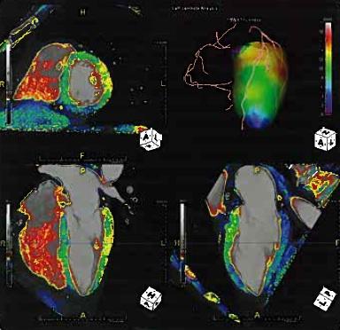
Advanced visualization software makers TeraRecon and Vital Images both showed FDA-cleared software that enables CT myocardial perfusion imaging. If this software can be proven equal to positron emission tomography (PET) or single photon emission computed tomography (SPECT) myocardial perfusion imaging, CT angiography will likely get a major boost and eliminate the need for a nuclear scan. Additional benefits of CT perfusion imaging would be increased diagnostic accuracy, because the detailed anatomy can be seen on the CT scan and culprit blockages can be visualized in the vessels from the same dataset.
Vital Images gained FDA-clearance for its CT Myocardial Analysis software in late November. The company is careful not to say "perfusion" imaging because of the limits of its FDA indication, but that is essentially what it does. It creates a color-coded map of contrast levels in the myocardium during the cardiac cycle. This map can be viewed as a bulls-eye target or as an overlay over a 3-D rendering of the heart. Areas of low contrast, thus poor perfusion, correspond with areas of ischemia or infarct.
TeraRecon's coronary review software is being marketed primarily to researchers so its abilities can be further validated. It can show color maps of blood perfusion in the myocardium during the cardiac cycle. The images look very similar to PET perfusion scans, but with much better resolution.
The software also can measure wall motion in the ventricles and display it as a series of colored bands, with each color representing a particular phase from the cardiac cycle. Areas with rainbow-colored bands evenly distributed show normal motion, while areas of little movement show the colors overlapping.
Nano Technology to Reduce CT Noise, Dose
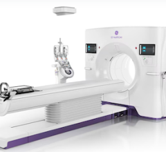
Siemens introduced its Somatom Definition Edge single-source CT system, a 510(k) investigational device, which uses new detector technology to reduce image noise and increase spatial resolution. It uses Siemens' new Stellar detector, which employs TrueSignal technology. It uses nano-electronic components to eliminate the traditional soldered circuit boards that surround today’s detectors. Noise is reduced by cutting the amount of electromagnetic activity in the circuits around the detector. It also integrates the electronic detector components into the photodiode.
These design changes are supposed to significantly reduce electronic noise and potentially improve the signal-to-noise ratio of the images. Siemens says a spatial resolution of up to 0.30 mm has been possible with the new detector. The company said the detector also helps in situations where the signal is greatly attenuated by adipose tissue, which may help generate better quality images in obese patients at lower doses.
CT Scan Dose Planning Software
Philips showed a conceptual work-in-progress of a CT dose treatment planning system that may help cut radiation to critical areas and only deliver dose to the anatomy of interest imaged. Similar to radiation treatment planning systems, Philips engineers showed conceptual CT planning maps of the patient’s anatomy that color-code critical, radiation-sensitive structures. After telling the computer what part of the anatomy is of interest for the scan, it would automatically figure how to image the patient with the lowest dose, possibly by modulating tube current, turning the tube on or off at different points of the scan or collimation.
If the technology could be made to work automatically and extremely fast, it may offer a new front for lowering CT dose.
Hands-Free Manipulation Images in the OR/Cath Lab
Siemens showed a work-in-progress system to allow OR surgeons or cath lab interventionalists to use mid-air hand motions to select a 3-D image on screen and allow hand motions to rotate the image. The technology would give the physician complete control of 3-D reference images without the need to touch anything or verbally instruct techs on how to manipulate the image remotely.
iPad-Like Ultrasound Controller May Replace Roller Ball
OEM Grayhill showed an ultrasound controller designed to replace the usual roller ball found on larger ultrasound systems. Instead, a large knob can be turned or pushed to select items and has a center pad that allows iPhone or iPad finger movements to zoom and select images. The concept will help make ultrasound consoles easier to sterilize and more intuitive to use.
Grayhill is an OEM ultrasound controller maker for some of the top ultrasound manufacturers.
Advanced Visualization for Patients
Advanced visualization company Vizua was formed 10 months ago and launched its U.S. presence at RSNA 2011. While it offers similar capabilities as other advanced visualization software vendors, it has a new business model that will likely gain attention. The company offers a low-cost cloud service to rapidly share images manipulated by advanced visualization that can be accessed easily by referring physicians or even patients, without downloading any software. The system uses intuitive, easy-to-use controls that patients can easily learn in a couple minutes. It has more advanced tools for physicians hidden in sub-menus.
The company plans to market its service to hospitals and imaging centers as a service enhancement. Currently, most referring physicians and the patients paying for the scans do not have access to advanced visualization-enhanced images.
TAVI Planning Software
In early November, the FDA cleared the first transcatheter aortic valve. Transcatheter aortic valve implantation (TAVI) technology is expected to revolutionize heart valve replacement with a minimally invasive procedure to replace open-heart surgery. However, it requires a good deal of planning, sizing and anatomical assessment of access routes using CT scans with manipulation with advanced visualization software. TeraRecon and Ziosoft both showed the first of what will likely be many TAVI planning software and tool set packages. These help automate manipulation of a CT dataset to quickly extract only the anatomy of interest and measurements, such as sizing of the aortic valve annulus and evaluation of clearance between the new valve and the right and left main coronary arteries.
Qi Imaging (formally Ziosoft) applied its deformable registration software to its TAVI package, allowing lifelike motion during the cardiac cycle to be applied. This allows for a more accurate assessment of the motion of annulus for better valve sizing.
3-D Mammography to Reduce False Positives
Fujifilm showed its Mammo Ascent 3-D mammography system that it hopes to commercialize in the next year. Users wear special glasses to view a set of two breast X-ray images shot a couple of degrees off from each other. The resulting true 3-D image enables easier evaluation of dense breast tissue, because the radiologist can differentiate between a mass and multiple thick tissue areas that are overlapping. It also shows whether several micro calcifications are in one mass or spaced over different tissue depths.
The company said one study using the system showed a 46 percent reduction in false positives and a 23 percent increase in sensitivity.
Trends Toward Simplicity and Cost-Effectiveness
Reflecting the mood of the economy and expectations from healthcare reform efforts, key messages from vendors at RSNA this year are lower cost, ease of use and increased efficiency. Adding to this, vendors also are expanding sales of their systems to developing countries, where there are severe shortages of trained technicians and physicians. Several vendors showed less expensive equipment options for ultrasound and CT systems. In some cases, vendors showcased systems with more automation but with fewer controls and buttons, making highly technical imaging equipment and software easier to use with better imaging results by less experienced staff.


 February 02, 2026


