GE's Vivid i
Although cardiac ultrasound has been around for many years, the technology is not static. Manufacturers are constantly looking for ways to improve the imaging, make the technology more accessible, affordable and now more portable.
The two most compelling changes in cardiac ultrasound are portability and improved imaging. Historically, one had to make a choice between portability and image quality. The newer machines have overcome the obstacles of combining clear images and small size.
Portability
Remember the days when the machines were so huge and cumbersome that the patients had to be transported to the machine? The machine took up the whole room. Today, the newer machines are amazingly small yet more powerful than their predecessors, making ultrasound technology truly point of care.
Portability in and of itself makes the ultrasound technology more available to the patient. According to Al Lojewski, global marketing manager, Cardiovascular Ultrasound at GE Healthcare, 60 percent of echos are being performed with portable devices. Cardiac ultrasound is finding its way into the ICU, operating room, emergency room and physician’s office. The idea proposed by Dr. John Gorcsan III in 2003 that portable ultrasound may be part of the bedside cardiology consult is now a reality.
The “portable” machines range from models that still require carts (but roll much easier than the 500-pound systems) to laptop-size devices that weigh as little as 5.5 pounds. The advantages of portable cardiac ultrasound are:
• Increased productivity as the machine can be carried patient to patient
• Can be used in areas where space is a precious commodity
• Easily transported to multiple settings, such as clinics
• Potential reduction in work injuries related to transporting heavy machinery
Improved Imaging
The utilization of computer technology has vastly improved ultrasound imaging since the early 1980s. The 2-D images that were standard for over 35 years have been improved to 3-D and 4-D. Images produced by some ultrasound packages rival the images seen in the multislice CT scanners, but without the hazards of X-ray exposure. Other technologies that have improved ultrasound imaging are:
• Power Doppler — said to be up to five times more sensitive than color Doppler
• Tissue Harmonic Imaging (THI ), which processes echoes at higher frequencies to reduce noise and artifact to better differentiate tissue, an important factor in using ultrasound on obese patients
• Real-time imaging
Variety of Technologies
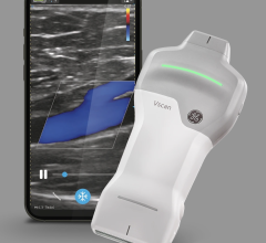
GE reports they are the only vendor who has products with the two newest advancements. In the extreme portability arena, their Vivid e and Vivid i systems are laptop sized. The
Vivid e weighs 10.1 pounds and is considered a completely portable cardiac echo. The Vivid i has a more expansive array of modalities depending on the package. The functions include but are not limited to stress echos, transesophageal echo and quantitative analysis of heart function. Their Vivid 7 Dimension '06 has 4-D capabilities and produces the images rivaling CT.
Zonare has trademarked the phrase “World's First Convertible Ultrasound” for its z.one platform. They have developed a new approach to ultrasound image acquisition called Zone Sonography. The acquisition of echo data in zones instead of narrow lines allows their system to process data very quickly.
According to Zonate literature, acquisition time for a representative frame is 5.2 msec versus 52 msec for conventional ultrasound. Because the system is software based both on the front and back ends, Zonare says its technology is flexible and easy to update, which reduces the cost of their product. The z.one converts from a full-featured cart-based product to a portable device that weighs only 5.5 pounds.
SonoSite manufactures an array of portable ultrasound devices including the MicroMaxx, a complete cardiac ultrasound machine weighing 7.7 pounds. SonoSite's ability to package the system so compactly is due to its proprietary Chip Fusion technology. If the idea of dropping a very expensive laptop makes you queasy, SonoSite originally produced the MicroMaxx for battlefield conditions and states it will function normally after a three-foot drop. The company offers a five-year warranty, which it says is unique in the industry.
Biosound Esaote's MyLab 30CV weighs less than 20 pounds and was introduced in 2004 as the first portable ultrasound with console performance. MyLab 30CV has various capabilities including multiplane TEE, integrated contrast acquisition and multiple analytics.
Siemens' cardiac ultrasound products range from the portable ACUSON Cypress weighing in at 19 pounds to the ACUSON Sequoia. Both products offer capabilities for TEE and intracardiac echo (ICE) with the use of its AcuNav ultrasound catheter. New technology recently released by Siemens such as syngo Auto Ejection Fraction technology is reported to calculate EF with 90 percent accuracy. The syngo Velocity Vector Imaging allows for assessment of myocardial mechanics and the syngo Quantitative Synch Tool is used for the assessment of ventricular dyssynchrony.
Toshiba offers a range of powerful cardiac ultrasound devices with a unique tool to reduce the incidence of work-related injury. Its iASSIST remote uses Bluetooth technology to allow the operator to work from a distance of up to 30 feet. The operator can define and activate procedure protocols with a push of a button so exam results can be duplicated even if they are complex. In addition, the remote reduces line-of-site limitations. Xario offers 4-D capability along with many sophisticated functions. Aplio CV offers applications measuring ejection fraction, myocardial tissue strain and tissue Doppler image quantification.
Many of the ultrasound products now offer various methods for data transfer and storage including wireless, CDROM, videotape and Digital Imaging and Communication in Medicine (DICOM) protocol. Some of the units have hard drive storage.
Intracardiac Echocardiography (ICE)
The list of technological advances in cardiac ultrasound would not be complete without the mention of intracardiac echocardiography (ICE). The need for intracardiac visualization arose with the advent of treating severe atrial fibrillation by percutaneous ablation as opposed to open Cox Maze procedures. Instead of blindly ablating tissue, the cardiologist can visualize in real time the targeted structures and the catheters they are using for the ablation. Without ICE, operators must use fluoroscopy to guide ablations, exposing both the operator and the patient to long exposure times due to the complexity of the procedure.
ICE can also be used to assess ventricular dyssynchrony and guide the placement of the left ventricular lead for resynchronization therapy.
As previously noted, Siemens currently offers ICE capability. In October 2006, GE Healthcare and St. Jude Medical announced a collaborative agreement to begin developing intracardiac echocardiography capability.
Boston Scientific offers an ultrasound imaging package via their iLab System with their Ultra ICE Catheter. Its system is available in two configurations, cart based or fully integrated into the electrophysiology system. Siemens’ ultrasound imaging system is also compatible with its IVUS and peripheral catheters.
Selecting Technology
There are multiple choices for cardiac ultrasound technology, and only a few have been mentioned here. Most of the manufacturers have a wide range of products depending on the buyer’s needs. When selecting the technology, here are key items to consider:
• What type of exams will it need to perform?
• How will it be reimbursed?
• Does it need to be mobile or very portable?
• What type of space is available?
• How easy is it to upgrade?
• Does your tech feel it's ergonomically safe?
• How durable is it if it will be moved frequently?
• Will education be required and is it available?
• What's the history of reliability?
• Will the vendor have a replacement unit available if yours is being repaired?
Conclusion
Cardiac ultrasound technology is changing at a rapid rate due to advances in computer software that allows for improved imaging in a smaller package. The ability to upgrade the technology may be as simple as new software or new microchip. Just like laptop computers, high tech doesn't necessarily mean high price. The increased functionality and portability can result in more efficient exams and better patient care.


 January 28, 2026


